ALPHAPetrol
Tartrate-Resistant Acid Phosphatase (TRAP) Stain Kit
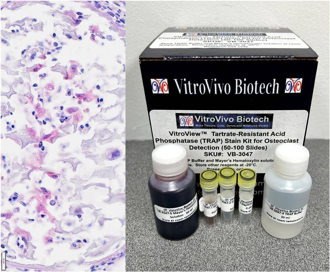
Description Tartrate-Resistant Acid Phosphatase (TRAP) is an acid phosphatase enzyme that remains active in the presence of tartrate. The enzyme hydrolyzes a chromogenic substrate, producing a colored reaction product at sites of TRAP activity. TRAP-positive cells typically appear as red, dark red, or purple red, depending on the kit formulation and counterstain used. Osteoclasts are generally identified as multinucleated TRAP-positive cells. This VitroView™ Tartrate-Resistant Acid Phosphatase (TRAP) Stain Kit is designed for histochemical identification of osteoclasts and other TRAP-positive cells in tissue sections and cell preparations. Key Advantages Dual-Modality Imaging: Enables both bright-field and fluorescence microscopy evaluation. Organic Mounting Compatible: Formulated with NewFuchsin to withstand alcohol dehydration and xylene clearing. High Yield Capacity: Single kit processes 50 to 100 slides efficiently. Versatile Sample Compatibility: Validated for cell smears, adherent cells, frozen sections, and paraffin sections. Streamlined Working Protocol: Micro-volume master mix preparation takes less than two minutes. Clear Cellular Contrast: Delivers distinct wine-red cytoplasm against light blue nuclei. Application Identification of osteoclasts in bone tissue Assessment of osteoclast differentiation in cell cultures Research applications involving bone remodeling and resorption Histological and cytochemical investigations Kit Contents VB-3047-1 NewFuchsin Solution 0.25 ml VB-3047-2 Sodium Nitrite Solution 0.25 ml VB-3047-3 Naphthol AS-BI Solution 0.25 ml VB-3047-4 TRAP Buffer 30 ml VB-3047-5 Mayer’s Hematoxylin Solution 30 ml Storage Store TRAP Buffer and Mayer’s Hematoxylin solution at room temperature away from light. Store other reagents at -20°C away from light. This kit is stable for at least 3 months. Procedure Sample Preparation: For Cell Smears (Blood / Bone Marrow): Prepare smears using fresh samples according to routine operations. Fix in 10% Neutral Buffered Formalin (NBF) for 15–30 minutes. Wash 3 times with distilled water. Proceed to Procedure Step 2. For Adherent Cells / Coverslips: Discard the culture medium completely. Wash 3–4 times with PBS. Fix in 10% Neutral Buffered Formalin (NBF) for 15–30 minutes. Wash 3 times with distilled water. Proceed to Procedure Step 2. For Frozen Sections: Warm the frozen sections to room temperature. Fix in 10% Neutral Buffered Formalin (NBF) for 15–30 minutes. Wash 3 times with distilled water. Proceed to Procedure Step 2. For Paraffin Sections: Deparaffinize sections in xylene (2 × 6 min), followed by rehydration in 100% ethanol (2 min), 95% ethanol (2 × 2 min), and 70% ethanol (2 min). Rinse in distilled water for 5 min and proceed to Procedure Step 2. Staining Prepare approximately 1.0 mL of New Fuchsin TRAP staining working solution, sufficient for staining 2-5 slides, as follows: In a microcentrifuge tube, combine 10 μL of NewFuchsin Solution with 10 μL of Sodium Nitrite Solution. Incubate the mixture at room temperature for 1 min. Add 1.0 mL of TRAP Buffer to the tube. Add 10 μL of Naphthol AS-BI Solution and mix thoroughly to prepare the working staining solution. Use hydrophobic barrier pen to draw a water-repellent circle around tissue sections or cells on the slide. Gently drop the working solution to cover the cells or tissue section on the glass slides and incubate at 37 °C in moisture chamber for 40–60 min. Drop Mayer’s Hematoxylin Solution onto the bone sections for 2–5 min; then wash the samples with running water for 15 min. Dehydration and mounting Dehydrate with 2 changes of 95% Ethanol and 2 changes of 100% Ethanol (2 minutes per change). Clear with 3 changes of xylene (5 minutes per change) Mount coverslip onto glass slide with Permount or some other suitable organic mounting medium. Observation: Bright-field Microscopy can be used to examine specimens. When observing fluorescence, use a rhodamine excitation filter (500–570 nm). Expected Results Expected Results under Bright-field Microscopy Osteoclast cytoplasm —-wine-red Nuclei———————– light blue Expected Results under Fluorescence Microscopy Osteoclast cytoplasm ——— red Positive Controls Mouse fetus whole sections (Spinal bone) Metaphyses or growth plates from juvenile mice or rats (3 to 6 weeks old) are the most common laboratory controls. Human giant cell tumor tissue sections References Nakamura A, et al (2025). Osteoclast visualization: Tartrate-resistant acid phosphatase activity staining using NewFuchsin compatible with non-aqueous mounting and tissue clearing. Methods X, 14: 103136 Luo G, et al (2025). Precision-targeting and dual silencing osteoclastogenesis and inflammatory pathways for the treatment of radiation-induced bone deterioration. Biomaterials Advances, 117: More Tartrate-Resistant Acid Phosphatase (TRAP) Staining Images Tartrate-Resistant Acid Phosphatase (TRAP) staining of a fetal mouse spine FFPE section. Multinucleated osteoclasts exhibit positive red staining, indicating TRAP activity. Original magnification, 10×. Tartrate-Resistant Acid Phosphatase (TRAP) staining of a fetal mouse spine FFPE section. Multinucleated osteoclasts exhibit positive red staining, indicating TRAP activity. Original magnification, 20×. Precautions: Handle reagents with care. Avoid contact with eyes, skin, or clothing. Do not ingest. Always wear gloves when handling chemicals. User Manual and Material Safety Data Sheet (MSDS) (PDF) VB-3047 User Manual VB-3047 MSDS
Related products
-
Hi-Vis Women's Workwear, Digital Printed 2Pc, Specialist Footwear, B/I Ovens
Select options This product has multiple variants. The options may be chosen on the product page
Notify me when the item is back in stock.